Lupine Publishers | Open access journal of Immunology and Infectious Diseases
Abstract
Chediak-Higashi syndrome (SCH) is a rare autosomal recessive genetic disorder characterized by oculo-cutaneous albinism,
immunodeficiency responsible for recurrent infections, predisposition to bleeding, And late neurological deterioration. The
pathognomonic sign is the presence of giant intracytoplasmic granules in most of the cells of the organism but often they are
identified in peripheral blood. In 85% of cases, CHS patients develop the accelerated phase characterized by an Hemophagocytic
Lymphohistiocytosis syndrome (HLH) responsible for a high mortality rate. The only current effective treatment of haematological
and immunological abnormalities remains allogeneic bone marrow transplantation, but without impact on skin manifestations or
subsequent neurological deterioration. It is all the more effective as it is performed before the onset of an HLH syndrome.
Introduction
Chediak-Higashi syndrome (SCH) is a rare autosomal
recessive genetic disorder characterized by oculo-cutaneous
albinism, immunodeficiency by cytotoxic activity of T lymphocytes
and natural killer cells responsible for recurrent infections,
predisposition to bleeding, And late neurological deterioration.
According to the International Union of Immunological Societies,
the SCH is a primary immunodeficiency by immune dysregulation
belonging to familial lymphohistiocytic haematophagocytosis
syndromes (HFH) with hypopigmentation [1]. The LYST-CHS1
(Lysosomal Trafficking Regulator Gene) gene was identified on the
long arm of chromosome 1 in 1q42-q43 [2,3]. This gene encodes
the CHS protein whose exact function remains imprecise. About
500 cases have been reported [4,5]. The diagnosis is oriented by the
clinical signs and facilitated by the study of the microscopic aspect
of the hair which highlights the presence of pigment aggregates;
But the pathognomonic sign of the disease is the presence of giant
intracytoplasmic granulations in most cells of the organism [6],
especially in peripheral blood or bone marrow. Approximately
85% of patients develop an acceleration phase characterized by
a syndrome of lymphohocytic hemophagocytosis (HLH), which
occurs during the first decade, rarely present at the onset of the
disease [7]; It is fatal in the absence of treatment [8]. Currently, the
only effective therapeutic option is bone marrow transplantation,
which improves haematological and immune abnormalities, but
does not prevent subsequent neurological deterioration. The
prognosis remains poor in the absence of a bone marrow transplant,
the death often occurring before the age of ten years.
Patients and Methods
They are four children followed in the pediatric department
of the CHU Mustapha of Algiers for Chediak-Higashi syndrome
between 2014 and 2017. The diagnosis was focused on the clinical
manifestations, the presence of giant intra-leukocyte granulations.
A genetic study in three children confirmed the diagnosis.
Results
These are three boys and one daughter with an average age of
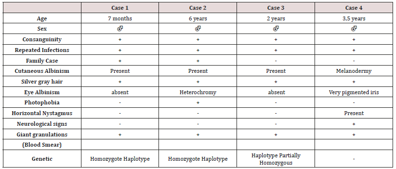
diagnosis of 3.2 years (7 months - 6 years). Consanguinity is found
in all cases as well as a family form (2 brothers). Three patients
have a history of repeated infections. Children who were vaccinated
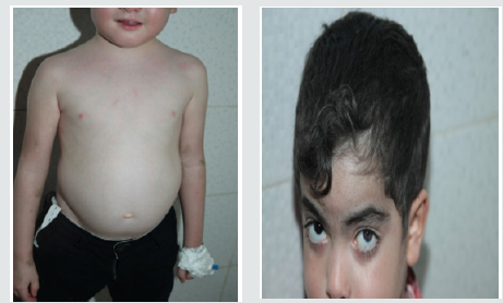
did not report any particular incidents. Clinically, oculocutaneous
albinism is present in 3 children (Figure 1) and melanoderma
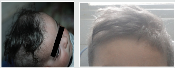
with a highly pigmented iris in 1 case (Table 1). Silver gray hair
is present in all patient (Figure 2). The peripheral blood smear
allowed to make the diagnosis by showing the intra-cytoplasmic
giant granulations in all the patients. Microscopic study of the
hair found deposits of melanin in irregular clods in the hair shaft
in favor of Chediak-Higashi syndrome (Figure 3). A syndrome of
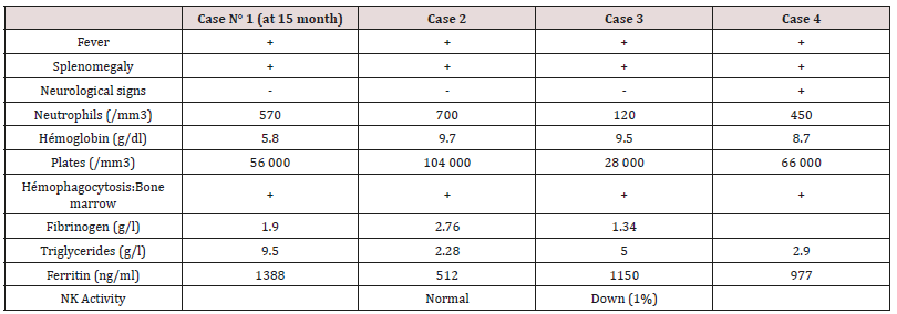
lymphohistiocyte hemophagocytosis (HLH) (Table 2) is present in
3 cases and then in one case 8 months after the diagnosis.
Figure 1: Hypopigmentation of the skin Clinically, oculocutaneous albinism is present in 3 children hypo pigmentation of the left iris

Figure 3: Deposition of melanin in the hair shaft Microscopic
study of the hair found deposits of melanin in irregular clods in
the hair shaft in favor of Chediak-Higashi syndrome

Melanoderma with a Highly Pigmented Iris in 1 Case
All patients with signs of lymphohistiocytic activation were
put on HLH 2004 protocol. The evolution is marked by a good
tolerance of the treatment. One child received an allogeneic marrow
transplant but died six months later. The remaining three patients
are still alive and clinically stable.
Discussion
Autosomal recessive genetic disease very rare, Chediak-Higashi
syndrome affects all races and all age groups. Less than 500 cases
have been reported [4,5]. Most often they are reported cases or
small series published. Prevalence is difficult to determine due to
reported cases more than once, and other unreported cases. The
largest published series (15 cases) was reported in Japan over a
period of ten years [9]. Inbreeding in all our patients is reported in
50 to 85% of cases [5]. The LYST / CHS1 gene of the disease encodes
the cytosolic CHS protein whose function remains imprecise. It
would play a role in the exocytosis of proteins from late
multivesicular endosomes. More than sixty mutations have been
reported in the literature (false sense, nonsense, deletion,
insertions) [10]. Mutations in the LYST / CHS1 gene lead to an
abnormal function of the CHS protein with an impairment of
intracytoplasmic transport, protein sequestration in giant
intracytoplasmic structures and blocking of the secretory function,
in particular that of leucocytes and melanocytes. Phenotype /
genotype correlations were reported, thus a deletion-type mutation
correlated with the early and fulminant onset of the acceleration
phase, while a missense-like mutation correlated with a better
prognosis with no acceleration phase and neurological deterioration
[11]. Clinical signs begin after birth, or before the age of 5 years.
Oculocutaneous albinism (AOC) is an important sign of diagnostic
orientation, present in three of our patients, characterized by
hypopigmentation which generally affects the skin, the hair and the
eyes. It is related to pathological aggregation and an unequal
distribution of melanosomes. The AOC may be present from birth,
and concern the three organs or some of them, total or partial or
even absent [12]. Sometimes hyperpigmentation as in one of our
patients can exceptionally be seen, delaying the diagnosis [13,14].
Most patients have photosensitivity. Some patients have an atypical
phenotype with an attenuated form, or the AOC is subtle or absent
and probably unknown [11,15]. Rarely other skin lesions are
observed as hyperhydrosis, erythema multiforme. The
hypopigmentation of the hair gives them a blonde, gray or white
color, often with a silvery or metallic luster. The eyes are blue in
color and hypo pigmentation of the iris may be associated with
decreased pigmentation of the retina, and ocular manifestations
such as photophobia, decreased visual acuity, nystagmus, and
strabismus Frequent and recurrent infections are common in
childhood. Often severe, they are related to a defect in T cell
cytotoxicity, NK function, and a decrease in the chemotactic and
bactericidal activity of the granulocytes [16,17]. Pyogenic infections
are the most frequent, especially in the skin, upper airways and
mucous membranes. The most frequently isolated germs are
staphylococcus aureus, β-hemolytic streptococci and
pneumococcus. The involvement of the oral cavity manifests in the
form of gingivitis, gingival haemorrhage, early falls of the teeth.
Mouth ulcers have been described. Periodontitis has been identified
as a manifestation of immune dysfunction. The bleeding tendency
in these patients is related to a deficiency in the storage pool of
dense granules and a defect in platelet aggregation. Haemorrhagic
manifestations are usually benign and usually do not require
treatment. The accelerated phase of the disease is the most
important and dangerous complication of the SCH. It is responsible
for a high rate of mortality within a few months [18]. It can occur at
any age but especially during the first decade (85%) [19]. Our
patients developed this accelerated phase before the age of 6 years.
Rarely, it is the first manifestation [7,20]. Its early onset is associated
with the existence of a deletion-like genetic mutation [11] and has
a collapsed or absent activity in cytotoxic LTs [21]. It is manifested
by a syndrome of lymphohystiocytic haematophagocytosis (HLH)
whose factors triggering this acceleration are not clear. The role of
EBV infection found in one of our patients was raised without this
relationship being established [22]. The diagnosis of accelerated
phase disease is based on the criteria of the Histiocytic Society
2004 [23]. Neurological manifestations occur in about 50% of cases
and may occur at any time in childhood or adulthood, they are
variable: peripheral neuropathy, coma, convulsions, ataxia,
cognitive disorders, impaired balance, movement abnormalities
and mental alterations. The bone marrow transplant does not
prevent their subsequent appearance [24]. The diagnosis of SCH
should be early, often done at around 6 years of age, but in about
25% of cases the diagnosis is delayed after age 10 [12] for our
patients. The average age at diagnosis was 3.2 years. It is suspected
on the clinical elements, facilitated by the microscopic study of the
hair which shows aggregates of melanin pigments found in all our
patients, this aspect allows the differential diagnosis with other
types of cutaneous hypopigmentation. But the pathognomonic sign
of the disease is the presence of giant intracytoplasmic granules in
most of the cells of the organism [6] but often they are identified in
peripheral blood as was the case of our patients or in the bone
marrow. The diagnosis is confirmed by a genetic test for the
mutation LYST. Antenatal diagnosis of the disease is possible in the
cells of the chorionic villi, amniotic fluid, leucocytes of the fetal cord
[25]. As with Chediak-Higashi disease, other genetic immune
deficiencies are accompanied by partial oculocutaneous albinism,
such as Griscelli’s disease and Hermansky-Pudlak syndrome. The
distinction can be made unambiguously by the different appearance
of pigment clusters in the hair sheath, much finer in the case of CHS,
and especially by the presence of giant intracytoplasmic granules
observed only in the CHS. However, in some cases of myeloid
leukemia, one can see giant granulations called pseudo Chediak-
Higashi anomaly [26]. The treatment of Chediak-Higashi disease is
multidisciplinary and is based on the management of complications
of the disease, treatment of the “accelerated phase” or HLH and
especially the transplantation of hematopoietic stem cells.
Symptomatic treatment of Chediak-Higashi disease is based on
effective antibiotic therapy against infections and transfusions of
blood derivatives to fight anemia and hemorrhagic complications.
Eye disorders should be corrected. The eyes and skin should be
protected from UV rays. Vaccinations are generally well tolerated as
was the case for our patients and must be done. Hygiene and oral
health care are Primordial. The occurrence of neurological
symptoms and their progression must be dealt with early enough
by a rehabilitation specialist. In the case of an acceleration phase
(HLH, a treatment combining corticosteroids, VP16, cyclosporin
and intrathecal injections of MTX (HLH 2004) [23] is introduced to
achieve remission, which occurs in 75% of cases [27], but relapses
are frequent and response to treatment decreases over time. When
transplantation is achieved, transplantation is recommended. In
SCH patients with HLH by EBV the addition of Rituximab could
improve treatment [28]. In the case of refractory HLH, another
therapeutic option including a monoclonal anti-CD52 antibody
(Alemtuzumab) [29] is possible as a second line treatment before
bone marrow transplantation. Allogeneic bone marrow
transplantation (BMT) is the only current effective treatment that
heals hematologic and immunological abnormalities, but has no
effect on oculocutaneous albinism or subsequent neurological
deterioration [9,24,30]. The pre-graft conditioning regimen
comprises a combination of etoposide, busulfan, cyclophosphamide
[31]. Reduced intensity of pre-graft conditioning with fludarabine,
melphalan, and alemtuzumab resulted in increased survival in
primary or family HLH with lower toxicity [32,33]. Bone marrow
transplantation is most effective when performed before the
accelerated phase occurs [31]. Patients with a profound decrease in
the cytotoxic function of T lymphocytes (CTL) have a high risk of
developing lymphocytic syndrome (HLH), so their screening may
be an indication for early marrow transplantation [21]. The overall
survival rate after marrow transplantation is 60-70% (30-32) The
prognosis remains poor in the absence of a bone marrow transplant,
death occurs frequently during the first decade by infections or
development of an accelerated phase HLH [34] About 10 percent of
patients who survive in early childhood will develop severe
neurological disorders in adolescence and early adulthood [35].
Conclusion
Chediak-Higashi syndrome is a rare disease, the diagnosis is
suspected in a child with oculocutaneous albinism with recurrent
infections. The majority of clinical forms are early “infantile” lethal
in the absence of treatment. A minority of patients present with an
“attenuated” form of the disease will survive after childhood but
develop an associated neuro-degerative disease. In all cases early
diagnosis should be posed by a simple examination, peripheral
blood smear which shows the presence of giant intracytoplasmic
granules pathognomonic of this condition. The only current effective
treatment of haematological and immunological abnormalities
remains allogeneic bone marrow transplantation, but without
impact on skin manifestations or subsequent neurological
deterioration. It is more effective when it is performed before
the onset of an HLH syndrome. In the event of the occurrence of
accelerated phase (HLH), a treatment according to the HLH 2004
protocol is instituted in order to obtain a remission before the
bone marrow transplant. The prognosis of infant form is poor,
with death occurring frequently in the first decade of life through
infections or development of HLH. The search for predictive factors
for the development of HLH may help in the indication of early bone
marrow transplantation.
For more Lupine Publishers Open Access
Journals Please visit our website:
For more open
access journal of Immunology & Infectious Diseases articles Please Click
Here:
To Know More About Open Access Publishers Please Click
on Lupine
Publishers
Follow on Linkedin : https://www.linkedin.com/company/lupinepublishers
Follow on Twitter : https://twitter.com/lupine_online


No comments:
Post a Comment