Lupine Publishers | LOJ Immunology and Infectious Diseases
Abstract
Keywords: Burukutu; Pig Hindgut; Lactic Acid Bacteria; Escherichia Coli Infection Body Weight; Haematology
Introduction
Materials and Methods
Study Area
The study was carried out in Samaru Campus of A.B.U, Zaria, Sabon Gari Local Government Area of Kaduna State and Located on Latitude (11°11´N, 07, 38´E, 686m above sea level) in the Northern Guinea Savannah with temperature ranging from 13.8° to 36.7° C. and an annual rainfall of 1092.8mm [11]. Agriculture in Zaria can be divided into two types: Rainfall type (from May to October) and irrigation farming in the dry season (from November to April). Dry season farming is the second most prevalent Agricultural activity in Zaria with vegetables being the common produce, but in some cases, fruits are sandwiched among cereal crops [12].Ethical Approval
Approval for the conduct of this research was obtained from the ethical clearance committee for animal use and care of Ahmadu Bello University Zaria; with the approval number: ABUCAUC/2016/005.Source of Experimental Animals
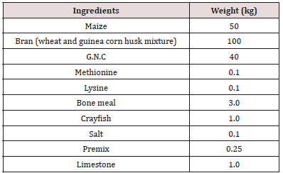
A total of 60 Large white breed of weaned pigs aged 42-60 days old with average body weight of 5.5 kg were sourced from breeders in Hayin Mallam, a small village (settlement) in Samaru, Sabon-Gari Local Government Area of Kaduna State and popularly known as ‘Pig-City’. Forty-eight (48) of them were sheltered in the piggery pen of the Division of Agricultural colleges Samaru, Zaria. Whilst the remaining 12 weaners which will serve as the control group, were housed separately in the Piggery pens of the Department of Veterinary Medicine, Faculty of Veterinary Medicine, A.B.U Samaru, Zaria, Kaduna State these separation was done, so as to avoid cross contamination of diseases. The Pigs were fed ad libitum with water and commercial Pig Weaner diet (Table 1) containing 21% crude protein and 3606 kcal ME/kg.The ingredients composing the feed were mixed thoroughly and bagged in 100kg grain bags to ease transportation and stored at room temperature in the feed store of the Department of Medicine, Faculty of Veterinary Medicine, Ahmadu Bello University, Zaria. Prior to the feeding with the experimental feed mixes, the following base-line parameters were determined for the experimental animals: For haematological parameters, white blood cells (WBC) especially neutrophils, lymphocytes, basophils and eosinophils were evaluated. These parameters were determined using the automatic blood analyzer (CELL-Dyn 3200, Abbott Lab, Abbott Park, IL). Blood Samples were centrifuged at 6.67g at 40C.
Body Weight
All the pigs in each group were weighted weekly, using a salter’s scale hung on a beam to support the animal. The following mix ratios of feed versus fermenters were used in mixing wet and dry feed forms for feeding of the experimental animals: 8 kilograms of the feed was weighed using a kitchen scale and poured into a pair of 10liters plastic buckets into which 0.8mls of the overnight culture of lactobacillus bacteria was added and thoroughly mixed. Then, 8 liters of distilled water was added to one of the buckets and stirred with a clean spatula while the feed-mix in the second bucket was left dry. This procedure was repeated for other fermenters. However, the control feed-mix was constituted without inclusion of any of the fermenters used for the other experimental feed-mixes.Experimental Design
The weaned pigs were tagged and randomly allocated into 5 groups of 6 pigs each with each group having a replicate (Table 2).Keys
WF+Lact = Wet Feed plus Lactobacillus bacteriaDF+Lact = Dried Feed plus Lactobacillus bacteria
Control BD (WF) = Basal diet in wet form
Control BD (DF) = Basal diet in dry form
Lac BKT = Lactic acid bacteria isolates from “burukutu”
Lac Pg = Lactic acid bacteria isolates from hindgut of pigs
Lac PSTAB®= Bacillus pumilis/Bacillus subtilis a (patented probiotic)
Lac PTAB®= Lactobacillus acidophilus a (patented probiotic)
Diets and Feeding Regimen
A grower diet was formulated for the experimental weaned pigs (Table 1). Then two dietary treatments were formulated with or without bacteria inclusions. 1. Dry feed (DF) supplied as meal and 2. Liquid feed supplied as wet form (WF). The liquid feed was prepared by mixing meal and water in a 1:4 ratio. The WF was prepared by mixing the feed with water in a 20 liters well labeled tank and agitated at 27°C (room temperature) before inoculating all the four treatment groups with lactobacillus from the different sources (BKT, Pig gut, Patented bacteria PSTAB and Patented bacteria PTAB). The control group was fed only the basal diet without LAB inclusion. For the dry feed, it was prepared by mixing the basal feed in four different 20 liters well labelled tanks which were agitated at 27°C (room temperature) before inoculating all the four treatment groups with lactobacillus from different sources (BKT, Pig gut, Patented bacteria PSTAB and Patented bacteria PTAB). The same procedure was applied for feed preparation like the first, but the only difference was that water was not added to the feed. For animals in the control group, only the basal diet was offered in dry and wet forms without LAB inclusion. The animals in all the groups were initially fed for a period of three weeks without the treatments (pre inoculation feeding). Then the various bacteria, isolates were added accordingly into their various rations as described by previous researchers [11,13]. The appropriate amounts of feed were consumed within approximately 30 min once daily in the [14]. according to their body weight so that the whole ration fresh water was also provided ad lib.Lactobacillus Enumeration and Isolation
After three weeks treatment period, faecal samples were collected from the rectum of all the animals in each treatment groups to ascertain the level of lactobacillus shed in the faeces. Approximately five (5)g of feces was collected from the rectum of each animal in the treatment group with a sterilized spatula, and with the animal properly restrained. The fecal samples were properly labelled and immediately transported to the Microbiology Laboratory of the Department of Veterinary Medicine, Ahmadu Bello University Zaria, and processed, using the following steps as described by [15]:a) Samples of feces were suspended in quarter-strength Ringer’s solution, homogenized with a classic blender (PBI International, Milan, Italy) and poured on plates containing prepared Rogosa agar (Oxoid Ltd.) which were properly labelled according to the treatment groups for total count of lactobacilli. After anaerobic incubation at 370 C for 24 hours, 10 colonies were randomly selected from plates containing the last sample dilution (10-8).
b) Isolates were cultivated in MRS broth (Oxoid Ltd.) at 370 C for 24 hours and re-streaked into MRS agar. The total Lactobacillus counts were carried out on MRS-agar plates. Numbers of colony forming units (CFU) was expressed as log10 CFU per gram.
c) Microscopy observation and Gram staining’s performed to confirm isolates.
Source of E. Coli for Infection of Pigs
Feces were collected from a diarrhoeic weaner pig in a piggery in Hayin Mallam in Samaru, placed into a sterile polythene bag and transported to the bacterial zoonoses laboratory of the Department of Veterinary Public Health and Preventive Medicine, Ahmadu Bello University Zaria. Ten (10g) of the feces was inoculated into 90mls of broth (Tryptone soya broth), incubated at 37ºC for 24hours for enrichment. A loop full of the broth culture was streaked on EMB agar medium and incubated for 24hours at 370 C. Colonies characteristic of E. coli (green metallic sheen) were tested using the method of characterization as mentioned in Bergey’s Manual of Systematic Bacteriology [16].Serotyping of Pathogenic E. Coli
A loop full of sterile normal saline was placed on a clean glass slide and a colony from the plate was picked and emulsified to form a homogenous mixture then a drop of the polyvalent phase3 sera (Oxoid, UK) was added and gently rocked for about five seconds until E.coli antigen were detected by agglutination reaction.Infection of The Weaners with E. Coli Isolate
The fed treatment LAB groups were challenged with E. coli. Each weaner received 6 mls (1x10¹⁰CFU·ml-1) orally, of the E. coli strain solution using a 10mls syringe that was held at the back of their oral cavities at 5.00 pm on day of infection according to the method described by [17].Faecal Sampling and Bacteriological Analysis of Challenged (Infected) Pigs
Faecal samples were collected from each weaned pig 15 hours after the challenge at 8.00am of the following day and serially diluted (0.1 in 9.9mls). Two tubes at 10-4 and 10-6 were selected and spread plated on EMB-agar (Oxoid, UK) for Enterobacteriaceae enumeration and incubated at 37°C for 24-48 h, as described by [5]. Number of colonies forming units (CFU) was expressed as log10 CFU per gram to obtain the number of colonies shed on a daily basis and this was done for a period of two weeks and results collated. Similarly, the control groups were also infected at the same time and observed for clinical signs of colibacillosis such as diarrhea, rough hair coat and lethargy and those showing such clinical signs were treated with Vetcotrim® bolus antibiotic (Kepro company UK, 5g/50kg per os).Results
Figure 1: Mean weekly body weight gain of weaned pigs
fed BKT lactobacillus isolates in wet and dry feeds.
A wet = wet feed + BKT bacteria Isolate
A dry = dry feed + BKT bacteria Isolate
E wet = wet feed only
E dry = dry feed only
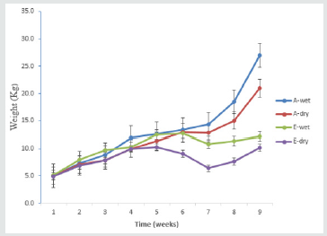
There was a significant (p<0.05) progressive increase in mean
weekly body weight in animals fed BKT bacteria inoculated wet
and dry form of feed when compared with the control animals fed
basal diet in wet and dried form. The group fed BKT in wet feed
form produced significantly heavier animals compared to those fed
dry feed with BKT bacteria. From week four, there was a gradual
increase in weight in all the treatment group and the control group
(p<0.05) but however, from week seven an exponential increase in
weight was observed in the treatment group and a marked decrease
in weight in the control group until week nine when the experiment
was terminated (Figure 1). The group fed PGUT bacteria isolates
showed a significant exponential (p<0.05) (Figure 2) increase in
weight which was as compared to the control group fed with basal
diet in both wet and dry form. However, the group fed PGUT bacteria
in wet form produced significantly (p<0.05) heavier animals as
compared to those fed inoculated dry feed. Also, there was a steady
weight gain between week four and seven in the group fed PGUT
bacteria in wet feed. And this increased significantly (p<0.05) from
week seven to nine, while the group fed dry feed with PGUT bacteria
isolate showed a gradual decrease in weight between week and six
but increased rapidly from week seven to nine. The control group
fed wet feed only showed a gradual increase in body weight from
week four to five then plateaued between week five and six and
gradually decreased till week nine while the control group fed dry
feed only, group started showing a decrease in body weight from
week four to seven before it increased steadily up to the ninth week
(Figure 2). The group fed with PSTAB bacteria in wet and dry form
of feed showed an exponential (p<0.05) increase in body weight as
compared to the control group fed basal diet in wet and dry form.
Though, animals in the treatment wet group where slightly heavier
than those in the treatment dry group. There was a gradual increase
in body weight in the treatment group from week four to seven
which later progressed until week nine. While the control group
showed a slow increase at week four which progressed steadily to
week nine though much lower than the treated groups (Figure 3).
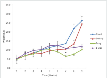
From week four, a gradual increase in body weight was observed
in all the treatment and the control groups but from week seven,
the treatment groups showed a significant (p<0.05) increase in
body weight as compared to the control. Also, for control animals,
a decrease in weight was observed from week five to week seven
with a gradual increase from seven to nine-week (Figure 4).
Figure 2: Mean weekly body weight gain of weaned pig
fed lactobacillus isolates from pig gut in wet and dry feed.
B wet = wet feed + PGUT bacteria Isolate
B dry = dry feed + PGUT bacteria Isolate
E wet = wet feed only
E dry = dry feed only
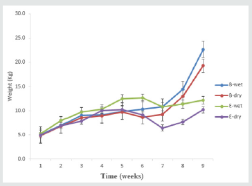

Figure 3: Mean weekly body weight gain of weaned pig
fed lactobacillus isolates containing Bacillus subtilis and
Bacillus pumilis in wet and dry feed.
C wet = wet feed + PSTAB bacterial Isolate
C dry = dry feed + PSTAB bacterial Isolate
E wet = wet feed only
E dry = dry feed only
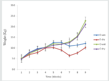

Figure 4: Mean weekly body weight gain of weaned
pigs fed lactobacillus acidiphilus formulated in pig food
supplement (tablet) in wet and dry forms.
D wet = wet feed + PTAB bacteria Isolate
D dry = dry feed + PTAB bacteria Isolate
E wet = wet feed only
E dry = dry feed only

Mean WBC Count
Effect of Probiotics on Haematological Parameters of Weaned Pigs Fed Actobacillus Fermented Feed
Mean Hemoglobin Value
Table 3: Effect of probiotics on the WBC values of weaned pigs for a period of nine weeks of pre feeding, feeding and challenged
with E. coli infection.
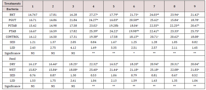

Table 4: Effect of probiotics on the hemogram (HgB) of weaned pigs for a period of nine weeks pre feeding, feeding and challenged
with E. coli infection.
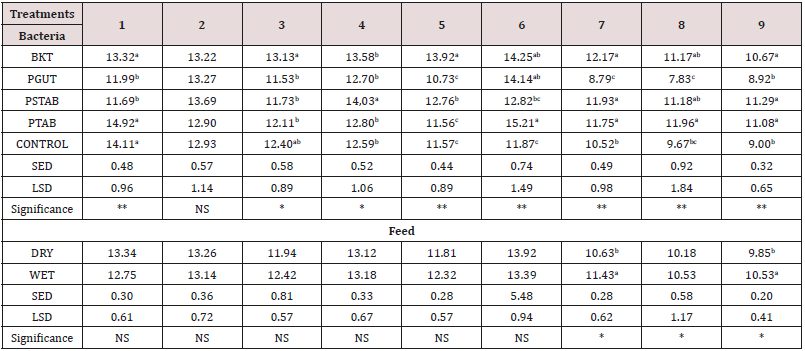
** = Significant at 1% and * at 5% level respectively. Mean within a
column of any set of treatment followed by different letter are
significantly different at 5%level while means followed by similar
letters in column are not significantly different
NS = Not Significant
SED = Standard deviation
BKT = Bacteria from burukutu
PGUT = Bacteria from pig intestine
PSTAB = Patented bacteria
PTAB, Patented bacteria

Table 5: Effect of probiotics on the hemogram (Neutrophils) of weaned pigs for a period of nine weeks pre feeding, feeding and
challenged with E. coli infection.
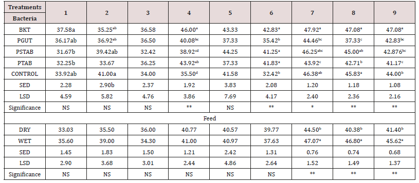
** = Significant at 1% and * at 5% level respectively. Mean within a
column of any set of treatment followed by different letter are
significantly different at 5%level while means followed by similar
letters in column are not significantly different
NS = Not Significant
SED = Standard deviation
BKT = Bacteria from burukutu
PGUT = Bacteria from pig intestine
PSTAB = Patented bacteria

Table 6: Effect of probiotics on the (Lymphocytes) of weaned pigs for a period of nine weeks pre feeding, feeding and challenged
with E. coli infection.
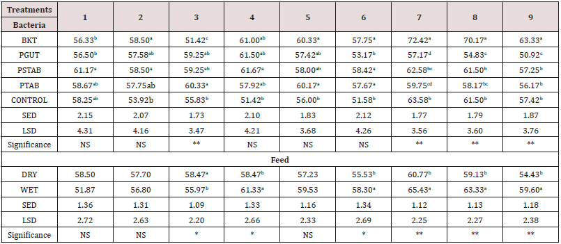
** = Significant at 1% and * at 5% level respectively. Mean within a
column of any set of treatment followed by different letter are
significantly different at 5%level while means followed by similar
letters in column are not significantly different NS = Not Significant.
SED = Standard deviation, BKT = Bacteria from burukutu, PGUT = Bacteria
from pig intestine, PSTAB = Patented bacteria, PTAB,
Patented bacteria

Discussion
Conclusion
The findings from this study have shown that: The addition of probiotics basal diets has promising effects on the performance of weaned piglets in terms of increasing body weight gain and improving the immune system thus reducing the severity of infection by pathogenic microorganisms. BKT inoculated feeds both in the wet and dried form, were found to be better than the patented bacteria as a probiotic as it increased the body weight as well as improved the bodies defence by increasing the immune system cells like WBC. Thus, locally sourced probiotics have like BKT and PGUT the potential of being patented for use in the Pig Industry in Nigeria.
Follow on Linkedin : https://www.linkedin.com/company/lupinepublishers
Follow on Twitter : https://twitter.com/lupine_online

No comments:
Post a Comment